The emergence of new animal models that mimic human disorders (neurodegeneratives diseases, tumour growth, neuropsychiatric disorders) enables new fundamental and therapeutical approaches of these diseases. The increasing number of studies on animal models implies the development of new imaging tools adapted to the particular constraints of small animal studies. To that aim, the past few years have seen the development of numerous high resolution PET systems having a sensitivity and a spatial resolution well suited to the small animal imaging. Nevertheless, although these systems have already and extensively demonstrated their interest in the biomedical imaging field, they suffer from a few drawbacks in the specific field of neuroscience (low temporal resolution, animal immobilization) that have stimulated the development of complementary approaches.
In that particular context, the main goal of the present project is to conceive a beta+ radiosensitive probe implantable in the brain of a rodent and able to measure in vivo and autonomously radioactive variations in a small and local tissue volume (a few mm3). Thus designed, this probe should allow the measurement of radiotracer kinetics on awake and freely moving animal without any physical connections, giving the possibility to combine simultaneously behavioural studies. The detection principle is based on the limited range of beta+ particles within biological tissues: this limited range allows the definition of a detection volume centered on the sensitive part of the probe. Beta+ particles emitted from distances further than their maximum range in biological tissues will be stopped before interacting with the probe and consequently will be not detected. This probe will be composed of Silicon pixels, connected to a dedicated integrated electronic board and finally, wireless linked to a data acquisition board including a signal receiver and set on a PC.
The project is carried out by a collaboration of 4 laboratories: the center of Imagerie et Modelisation en Neurobiologie et Cancerologie (IMNC, UMR8165) where a first probe prototype was previously developed, the Centre de Physique des Particules de Marseille (CPPM, UMR6550) that previously developed numerous two-dimensional silicon pixelized detectors for particle physics which present a number of technical advantages to conceive a telemetric probe, CERMEP, one of the main French PET center, which has extensively validated the first probe prototype in the context of neuropharmacological experiments, and the center of Neurobiologie de l’apprentissage, de la mémoire et du comportement (NAMC, UMR 8620), that has a great expertise in animal behaviour and in the design of behavioural studies.
The project is divided in three main parts :
The first is mainly instrumental and devoted to the production of miniaturized coated probe prototypes and the design of a specific and dedicated electronic. This part includes different steps of physical assessments (beta et gamma characterization and electronic validation), the design of the electronic block supporting the autonomous battery and its packaging (backpack) and finally, the acquisition system .
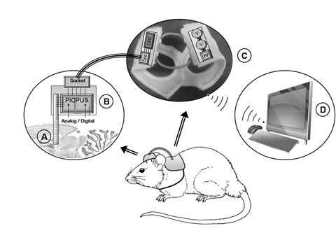
Design of the PIXSIC system. The system is built to be autonomous and easily worn by the animal: the detector (A) is sealed to the head of the animal with its front-end electronics (B). The other electronic components are deported on his back (C), and the signal is radio transmitted (D).
The second is more methodological and includes part of biological and physical approaches. It is first dedicated to the study of the brain injury and recovery after probe implantation and the putative consequence on the probe measurements during time. This part also includes the feasibility to realize with the pixelized probe a cartographic study of the radioactive distribution in tissue.
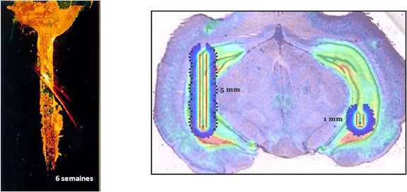
Left : illustration of glial scare following a probe implantation in rat brain.
Right : Simulation of mono and multi-pixel probes and their corresponding detection volumes after implantation in the rat hippocampus and injection of 18F-MPPF, a 5HT1A PET radiotracer.
![]()
The third is biological and especially dedicated to the validation of the set up in a biological context including first a comparison between anesthetized and awake animals and second, a measurement combining simultaneous radioactive measurements and behavioural studies.
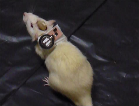
Initial behavioral test performed on rat using a first design of PIXSIC électronics and after chronic implantation of a 200 µm thick probe..
![]()
Globally, the project has to validate the interest of using silicon pixels to achieve the pointed biological studies in term of sensitivity and detection volume. A particular effort is focused on the probe temporal resolution which should allow to perform accurate kinetics measurements of radiotracer binding in the area of interest. Finally, the project has to show for the first time and in different biological contexts the ability of the probe to achieve quantitative measurements of a radiotracer on awake and fully freely moving animal. A patent focused on the probe principle allowing the freely movements of the animal during experiments has been deposited and at present under international extension demand (PCT/FR2008/001407).